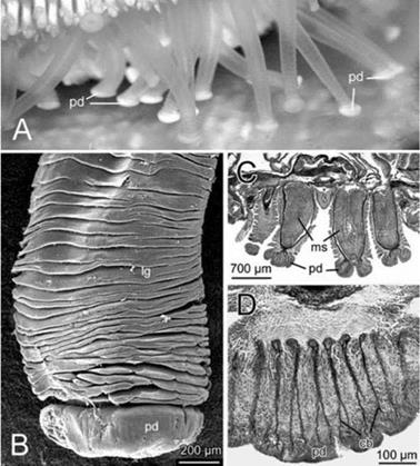
Echinodermata (starfish, see urchins, sea cucumbers) bear tube feet or podia that are used to adhere to and release from substrates by chemical interactions (Figure 8.85).
|
Figure 8.85 Podium of the starfish, Asterias rubens. (A) Starfish adhering to glass; (B) side aspect as viewed in the scanning electron microscope; (C, D) longitudinal sections through the podium. cb = collagen bundles; lg = leg; ms = muscles; pd = podium. |
This mechanism allows them to move [99,100] and burrow on substrates. The podia are external appendages of the hydraulic (ambulacral) system, and are driven partly by muscles and partly by hydraulic pressure [101]. The following types of podia have been identified: descending, ramified, knob ending, lamellate and digitate [102].
The histological structure of podia is relatively constant, there being four tissue layers: an inner and outer epithelium, with a connective tissue layer and a nerve plexus between them. The connective tissue layer is made from an amorphous substance that encloses bundles of collagen fibers and various types of cell. The layer consists of a diffuse external layer and a more compact internal layer within which the collagen fibers are arranged in circular fashion. In starfish and sea cucumbers, the external connective layer may contain one or several types of embedded calcareous needle (spicules) [103, 104]. Externally, a multilayered (three to five sublayers) cuticle covers the epidermis, consisting of fibrous and, sometimes, granular material [102].
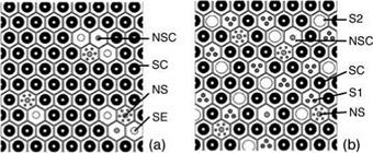
Only the distal parts of the tube feet (knobs, discs) are involved in attachment. Adhesive organs operate according to mechanical and chemical mechanisms; the podia possess an adhesive system with two types of secretory cell — those that release an adhesive secretion and those that release a de-adhesive secretion. In the podia epidermis there are three main types of specialized cell: secretory, neurosecretory — like, and sensory. In the handling podia the cells are always closely associated, forming sensory-secretory complexes (Figure 8.85) [102], but in the locomotory podia three cell types form a homogeneous cellular layer together with support cells (Figure 8.86). Such a cellular organization of the adhesive surface correlates well with the functions of the podia; a large adhesive area correspondingly provides a stronger attachment during locomotion, whereas discrete adhesive areas seem to be more efficient for the handling of small substrate particles.
Adhesive and de-adhesive secretions are produced by secretory and neurosecretory-like cells, respectively. In most species, the secretory cells contain acid mucopolysaccharides, although in several species proteins rather than mucopolysaccharides have been revealed in these cells. The third group of species possesses both
|
Figure 8.86 Schematics of transverse sections through adhesive areas of echinoderm podia. (a) Handling podia of Asteronyxloveni (Ophiuroidea); (b) locomotory podia of Holothuria forskali (Holothuroidea). NS = neurosecretory-like cells; NSC = sensory cells; S1, S2 and SE = secretory cells; SC = support cell. |
substances. Presumably, different functions of podia require different adhesive strengths and, therefore, adhesive secretions with different composition. Thus, a coarse molecular model for adhesion in echinoderms is a protein-polysaccharide complex, the composition of which varies from one taxon to another [102].
Interestingly, neurosecretory-like cells, which secrete de-adhesive substances, do not stain with any of the classical histochemical dyes. Although the mechanism of detachment is not fully understood, two hypotheses for its explanation have been proposed:
• De-adhesive material interferes with the electrostatic bonds between the adhesive layer and external layer of the cuticle.
• The material works as an enzyme, releasing an external layer of the cuticle from the underlying cuticular layers.
The mechanical mechanism of attachment is restricted to the sucker-mediated attachment. This hypothesis presumes that descending podia can deform the disc shape into a form ofsucker that is additionally supplemented by chemical adhesion.
The levator muscle in the podial disc is a probable candidate for suction generation. Recent experimental data on the mechanics of the tube feet show very soft properties of their terminal ends.
 16 января, 2016
  Pokraskin

 Опубликовано в рубрике