Marine bivalve mollusks are probably the most studied adhesive marine organisms. Their adhesive capacity is advantageous, because it allows mollusks to exploit nutrient-rich turbulent zones of the sea.
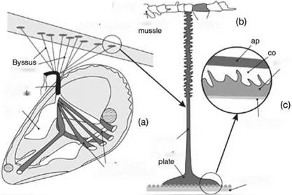
Oysters are permanently fixed to the substrate by secretion of cement glands (see Figure 8.82), whereas byssated mollusks adhere but are not necessarily permanently attached. They are able to abandon an old attachment site and move to a new one. The byssus is an extracellular structure consisting of a thread (0.05-0.20 mm in diameter and 2-4 cm long) attached to the animal at one end and to a substrate at the other (Figure 8.84) [95, 96]. An additional advantage of the byssus are its mechanical properties, allowing better damping of the wave impact.
|
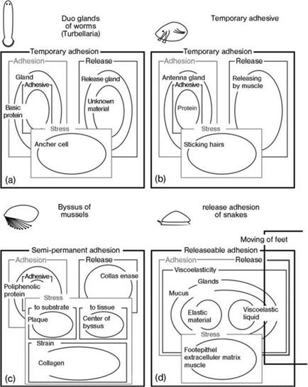
Figure 8.83 Schematic comparison of functional elements of adhesive organs in atubellarian (a), barnacle (b), mussel (c) and gastropod (d). The functions are indicated by boxes, and the structures which perform them as ovals. |
The foot produces the byssus, and the attachment to the animal is at the ventral base of the foot. The threads end at the stem, a root-like process embedded deeply in the base of the foot. The distal ends of the threads are attached to oval plaques (2.6 mm long and 2.0 mm wide).
The average attachment strength of the whole composition of threads of an animal ranges from 10-12 N in Mytilus edulisto 90 N in Septiferbifurcatus [95]. However, such
 With draw
muscle of byssus
Figure 8.84 The mussel byssus is a bundle of threads consisting of extracellular material. By drawing itself up on these tendinous threads, the mussel can control the tension of its attachment.
The distal tips ofthe threads end in adhesive pads called plaques.
(a) Schematic view of a mussel suspended by a byssus;
(b) schematic of the byssus; (c) distribution of mucosubstance (m), collagen (co) and polyphenolic protein (pp).
ap = adhesive pad.
a force is summed over all structural elements securing animals against external forces. It includes an adhesive bond of the plaque to the substrate, the thread attachment strength to the mussel, and the tensile strength of the threads. Usually, the breaking strength of threads is greater than that of the attachment sites. The proximal region and adhesive plaque are common sites of structural failure [97].
Byssal threads consist ofhighly ordered crystalline polymers (collagen and b-type protein), with the collagen fibers packed inside a b-type protein sheath [98]. Occasionally, the byssal threads consist of striated fibers.
Five groups of cells or glands participate in byssus production. In most species, the byssus consists of at least four essential components: acid mucopolysaccharides; adhesive protein; fibrous protein; and an oxidative enzyme, polyphenoloxidase. Many chemical details of byssus secretion remain unknown.
The sequence of events during the formation of the attachment plaque has been previously reviewed [95]:
1. With a contact angle of about 6°, a dispersion consisting of phenol granules and a mucoid substance is secreted onto the substrate surface. This dispersion is presumably mixed and applied to the surface in films 50 nm thick by use of the paddle-shaped cilia. Such a mixture results in a polyphenolic protein forming the continuous bonding phase.
2. The secretion becomes increasingly heterogeneous, with collagen added via the longitudinal ducts as the third component. The architecture of this heterogeneous material is cellular (trabecular): spaces between the trabeculae of phenolic material are filled with mucoid secretion. The collagen interacts with phenolic material and penetrates the trabecular architecture to a depth of about 3 mm.
3. The outer coverage of the thread and plaque is a dense substance, probably b-type protein.
As a result of these events, the byssal attachment plaque joins two dissimilar materials, a surface and the fibrous material of the threads.
 16 января, 2016
  Pokraskin
 Опубликовано в рубрике