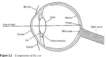
The sensation of colour that we experience arises from the interpretation by the brain of the signals that it receives via the optic nerve from the eye in response to stimulation by light. This section contains a brief description of the components of the eye and an outline of how each of these contributes to the mechanism by which we observe colours. Figure 2.2 shows a cross-section diagram of the eye, indicating some of the more important components.
The eye is enclosed in a white casing known as the sclera, or colloquially as the ‘white of the eye’. The retina is the photosensitive component and is located at the rear of the eye. It is here that the image is formed by the focusing system. Light enters the eye through the cornea, a transparent section of the sclera, which is kept moist and free from dust by the tear ducts and by blinking of the eyelids. The light passes through a transparent flexible lens, the shape of which is determined by muscular control, and which acts to form an inverted image on the retina. The light control mechanism involves the iris, an annular shaped opaque layer, the inner diameter of which is controlled by the contraction and expansion of a set
|
|
of circular and radial muscles. The aperture formed by the iris is termed the pupil. Light passes into the eye through the pupil, which normally appears black since little of the light entering the eye is reflected back. The diameter of the pupil is small under high illumination, but expands when illumination is low to allow more light to enter.
The retina owes its photosensitivity to a mosaic of light sensitive cells known as rods and cones, which derive their names from their physical shape. There are about 6 million cone cells, 120 million rod cells and 1 million nerve fibres distributed across the retina. It is the rods and cones that translate the optical image into a pattern of nerve activity that is transmitted to the brain by the fibres in the optic nerve. At low levels of illumination only the rod cells are active and a type of vision known as scotopic vision operates, while at medium and high illumination levels only the cone cells are active, and this is gives rise to photopic vision. Only one type of rod-shaped cell is present in the eye. The rods provide, essentially, a monochromatic view of the world, allowing perception only of lightness and darkness. The sensitivity of rods to light depends on the presence of a photosensitive pigment known as rhodopsin, which consists chemically of the carotenoid retinal bonded to the protein opsin. Rho — dopsin is continuously generated in the eye and is also destroyed by bleaching on exposure to light. At low levels of illumination (night or dark-adapted vision), this rate of bleaching is low and thus there is sufficient rhodopsin present for the rods to be sensitive to the small amounts of light. At high levels of illumination, however, the rate of bleaching is high so that only a small amount of rhodopsin is present and the rods consequently have low sensitivity to light. At these higher levels of illumination, it is only the cone cells that are sensitive. The cones provide us with full colour vision as well as the ability to perceive lightness and darkness. The sensitivity of cones to light depends on the presence of the photosensitive pigment iodopsin, which is retained up to high levels of illumination. Thus, in normal daylight when the rods are inactive, vision is provided virtually entirely by the response of the cone cells. Under ideal conditions, a normal observer can distinguish about 10 million separate colours. Three separate types of cone cells have been identified in the eye and our ability to distinguish colours is associated with the fact that each of the three types is sensitive to light of a particular range of wavelengths. The three types of cone cell have been classified as long, medium and short, corresponding to the wavelength of maximum response of each type. Short cones are most sensitive to blue light, the maximum response being at a wavelength of about 440 nm. Medium cones are most sensitive to green light, the maximum response being at about wavelength of about 545 nm. Long cones are most sensitive to red light, the maximum response being at about 585 nm. The specific colour sensation perceived by the eye is governed by the response of these three types of cone cells to the particular wavelength profile with which they are interacting.
 22 августа, 2015
  Pokraskin
 Опубликовано в рубрике