Since wood fracture usually dominates the performance of well-made joints, it is worthwhile before focusing on the bonded joint and the influence of the adhesive to examine how wood itself fractures. At the molecular level, Porter [19] found that wood fractures in the amorphous, water-accessible regions of the cell wall rather than in the crystalline regions. These regions are also most susceptible to change as a result of varying temperature, moisture content, and chemicals. At the microscopic level, wood fractures in different locations depending on the type of cell, direction of load, temperature, moisture content, speed of test, grain angle, wood pH, and aging.
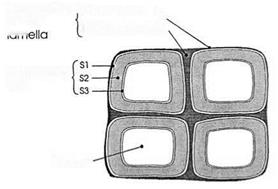
Anatomical features such as the S1, S2, and S3 layers of the cell wall (Fig. 2) are especially important in the fracture of wood and wood-adhesive joints. There are three general types of fracture at the microscopic level [20]: transwall, intrawall, and intercellular
 Compound Гprimary wall x
middle…… , „
I. —Middle lamella
Secondary
wall
Lumen
Figure 2 Transverse cross section of four wood cells showing the compound middle lamella joining them, the three layers (S1, S2, and S3) of the secondary wall, and the cell lumen.
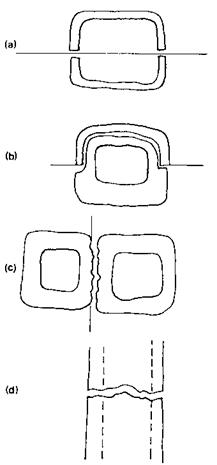
(Fig. 3). Transwall cracks may be parallel to the longitudinal cell axis (Fig. 3a) or transverse (Fig. 3d), but in either case the cell lumen is exposed. Transwall fractures are common in thin-walled cells such as softwood earlywood tracheids, hardwood vessels, and parenchyma cells. Longitudinal transwall fracture of thick-walled latewood cells is unusual. When such fracture occurs, it is extremely fibrous and is called fine-fiber failure [21]. Transverse transwall fracture (Fig. 3d) is rare in thick-walled cells (such as hardwood fibers and softwood latewood tracheids) as a result of their great tensile strength parallel to the cell axis. Such fracture does occur in compression wood of softwoods and at the tips of splinters in tough wood. These thick-wall cells are more likely to produce a diagonal combined shear and tension transwall fracture following the helical angle of the S2 layer microfibrils (not pictured). This is the manner in which a crack grows across the grain in tough wood. Intrawall fracture (Fig. 3b) is also very common in thick-walled cells. An intrawall crack travels within the cell wall, leaving the cell lumen intact.
Intrawall fracture initiates at the discontinuities between the layers of the secondary wall (Fig. 2). The cell wall consists of microfibrils of cellulose helically wound around the longitudinal cell axis. The cell wall layers are differentiated by the angles of the microfibrils in each layer. The microfibrils in the outermost (S1) and innermost (S3) layers are wound at a large angle around the longitudinal axis of the cell. The microfibrils in the S2 layer sandwiched between the S1 and S3 layers are wound at a small angle around the longitudinal cell axis. The transition between these layers is often gradual, yet it still presents a material discontinuity. Mark [22,23] clearly pinpointed the S1-S2 interphase as the site of crack initiation in the fracture of solid wood. Intercellular cracks (Fig. 3c) travel in the compound middle lamella (CML), leaving the secondary wall and cell lumen intact.
Investigators have shown the preferential fracture of wood at various cell wall interfaces, depending on the temperature at fracture. Woodward [24] found fracture predominately in the S1 layer in the range from 20 to 77° C. At the lower end of the scale, the crack path jumped back and forth across the middle lamella from the S1 layer of one cell to the S1 layer of a contiguous cell. At the higher temperature, the crack tended to stay within the S1 of a given cell from one end to the other. Furthermore, fractures of the lignin-rich CML are rare at normal temperatures, but they are likely to occur under hot, wet conditions [20].
On a larger scale, the type of fracture varies with the density of the tissue through which a crack is growing. Fracture in the longitudinal-tangential (LT) plane is dominated
|
|
Figure 3 Schematic diagrams of fracture modes in wood: (a) longitudinal transwall; (b) intrawall; (c) intercellular; (d) transverse transwall.
by longitudinal transwall fracture of the first-formed earlywood cells. A mixture of transwall and intrawall fracture is common in the longitudinal-radial (LR) and planes intermediate to the LR and LT planes as a result of alternating high — and low-density bands of the earlywood and latewood cells. Fracture patterns similar to those described for wood have been observed in solid wood joints and in wood particles bonded with droplets of adhesive [25,26].
 11 июля, 2015
  Malyar
 Опубликовано в рубрике